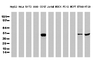
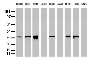
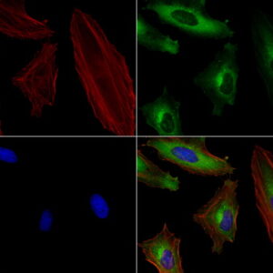
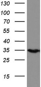
Purified P53 (TP53) mouse monoclonal antibody, clone UMAB62, 100 uL/ 30 uL
R$12.423,26
| Imunógeno | Proteína recombinante humana de comprimento total de TP53 humano (NP_000537) produzida na célula HEK293T. |
| Aplicações | IHC 1:50~100, |
| Aplicações2 | IHC |
| Resumo | A proteína codificada por este gene é um membro da família STAT de fatores de transcrição. Em resposta a citocinas e fatores de crescimento, os membros da família STAT são fosforilados pelas quinases associadas ao receptor, e então formam homo ou heterodímeros que se translocam para o núcleo da célula onde atuam como ativadores de transcrição. Essa proteína é ativada e medeia as respostas de muitos ligantes celulares, como IL2, IL3, IL7 GM-CSF, eritropoietina, trombopoietina e diferentes hormônios de crescimento. A ativação desta proteína no mieloma e linfoma associada à fusão do gene TEL/JAK2 é independente do estímulo celular e tem se mostrado essencial para a tumorigênese. A contraparte de camundongo deste gene induz a expressão de BCL2L1/BCL-X(L), o que sugere a função antiapoptótica deste gene nas células. |
| Formulação | PBS (pH 7.3) contendo 1% BSA, 50% glicerol e 0.02% azida sódica. |
| Purificação | Purificado a partir de fluidos de ascite de camundongo por cromatografia de afinidade |
| Isotipo | IgG1 |
| Reatividade | Humano |
| Hospedeiro | Camundongo |
| Tamanho | 43.5 kDa |
| Tipo | UltraMAB |
| Concentração | 0.74mg/ml |
Disponível por encomenda
Prediction model of lymphovascular invasion based on clinicopathological factors in Chinese patients with invasive breast cancer
Sandi Shen, Guihua Wu, …, Haibo Zhou
“2.3 ImmunohistochemistryEach TMA block was cut into 2.5-μm thick sections that were stained with the monoclonal antibodies ER, PR, HER-2, Ki67, EGFR, VEGF, E-cadherin, and P53. All the antibodies were ready-to-use from OriGene. A positive control was taken from a breast cancer tissue sample with positive IHC staining results, and a negative control was taken from a paraffin-embedded breast cancer tissue sample that had not been submitted for incubation with primary antibody. The alkaline phospha”
Produtos relacionados
-
 R$12.423,26
Imunógeno Fragmento de proteína recombinante humana correspondente aos aminoácidos 1544-1830 de DOCK2 humano (NP_004937) produzido em E. coli. Aplicações WB 1:2000, IHC 1:150, Aplicações2 WB,…
-
 R$12.423,26
Imunógeno Proteína recombinante humana de comprimento total de JUN humano (NP_002219) produzida em E. coli. Aplicações IHC 1:100, IF 1:100 Aplicações2 IHC Resumo Este gene…
-
 R$12.423,26
Imunógeno Fragmento recombinante expresso em E. coli correspondendo aos aminoácidos 240-390 de CK19 humana Aplicações WB 1:500~1000, IHC 1:50, IF 1:100 Aplicações2 WB, IHC Resumo A queratina 19 é um membro da família da queratina. As queratinas são proteínas de filamentos intermediários responsáveis pela integridade estrutural das células epiteliais e são subdivididas em citoqueratinas e queratinas capilares. As citoqueratinas do tipo I consistem em proteínas ácidas que estão dispostas em pares de cadeias de queratina heterotípicas. Ao contrário de seus parentes relacionados, esta menor citoqueratina ácida conhecida não está emparelhada com uma citoqueratina básica nas células epiteliais. É expresso especificamente na periderme, a camada superficial transitória que envolve a epiderme em desenvolvimento. Formulação PBS (pH 7.3) contendo 1% BSA, 50% glicerol e 0.02% azida sódica. Purificação Purificado a partir de fluidos de ascite de camundongo por cromatografia de afinidade Isotipo IgG1 Reatividade Humano Hospedeiro Camundongo Tamanho 43.9 kDa Tipo UltraMAB Concentração 0.5-1.0 mg/ml -
 R$12.423,26
Imunógeno Fragmento de proteína recombinante humana correspondente aos aminoácidos 57-314 da HADH humana (NP_005318) produzida em E. coli. Aplicações WB 1:2000, IHC 1:100~200, Aplicações2 WB,…