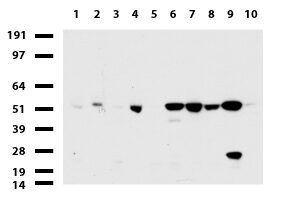
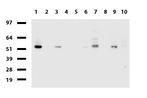
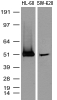
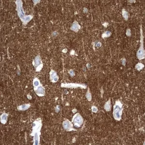
NCAM1 mouse monoclonal antibody, clone UMAB83, 100 uL/ 30 uL
R$12.423,26
| Imunógeno | Fragmento de proteína recombinante humana correspondente aos aminoácidos 20-718 de NCAM1 humano (NP_851996) produzido em E. coli. |
| Aplicações | IHC 1:100, |
| Aplicações2 | IHC |
| Resumo | Este gene codifica uma proteína de adesão celular que é membro da superfamília das imunoglobulinas. A proteína codificada está envolvida em interações célula a célula, bem como interações célula-matriz durante o desenvolvimento e diferenciação. A proteína codificada mostrou estar envolvida no desenvolvimento do sistema nervoso e para células envolvidas na expansão de células T e células dendríticas que desempenham um papel importante na vigilância imunológica. O splicing alternativo resulta em múltiplas variantes de transcrição. |
| Formulação | PBS (pH 7.3) contendo 1% BSA, 50% glicerol e 0.02% sódio azido. |
| Purificação | Purificado a partir de fluidos de ascite de camundongo por cromatografia de afinidade |
| Isotipo | IgG1 |
| Reatividade | Humano |
| Hospedeiro | Camundongo |
| Tamanho | 94.4 kDa |
| Tipo | UltraMAB |
| Concentração | 1.00mg/ml |
” followed by an additional wash with PBS for 5 min. Tissues were subsequently probed with primary antibodies for 1 h at room temperature. The following primary antibodies were used: Anti-CD3 (cat. no. LN10; 1: 50–100 depending on the tissue samples; OriGene Technologies, Inc.), anti-CD56 (cat. no. UMAB83; 1:100; OriGene Technologies, Inc.), anti-CD20 (cat. no. L26; 1:100; Dako; Agilent Technologies, Inc.), anti-TIA-1 (cat. no. 2G9A10F5; 1:100; OriGene Technologies, Inc.), anti-granzyme B (cat. no”
“Histopathology and immunohistochemistrySurgical samples were collected and embedded with paraffin for histological and immunohistochemical analyses. Immunohistochemical analyses for synaptophysin (SYN) (rabbit monoclonal antibody, OriGene, Rockville, MD, USA), chromogranin A (CGA; mouse monoclonal antibody; OriGene), cluster of differentiation (CD56; mouse monoclonal antibody; OriGene), cytokeratin (CK; mouse monoclonal antibody; OriGene), vimentin (VIM; mouse monoclonal antibody; OriGene), S-10”.
” incubation. The primary antibodies used were anti-HA (Roche) as well as, where available, primary antibodies against the native human genes. The primary antibody concentration used was 1 μg/μl for the protein specific antibodies (anti-Ncam1, UMAB83, Origene and anti-Nsf, ab16681, Abcam) and a 1:1000 dilution for the mouse anti-HA. Membranes were incubated with the secondary antibody (GE Healthcare) at a 1:5000 concentration for one hour, followed by signal detection using the Amersham ECL system”.
Produtos relacionados
-
R$12.423,26Adicionar ao carrinho
Imunógeno Fragmento de proteína recombinante humana correspondente aos aminoácidos 311-482 de TYMP humano (NP_001944) produzido em E. coli. Aplicações IHC 1:100, Aplicações2 IHC Resumo Este…
-
R$12.423,26Adicionar ao carrinho
Imunógeno Fragmento de proteína recombinante humana correspondente aos aminoácidos 323-547 de DDX56 humano (NP_061955) produzido em E. coli. Aplicações WB 1:2000, IHC 1:100~200, Aplicações2 WB,…
-
R$12.423,26Adicionar ao carrinho
Linfócitos e Macrófagos Infiltradores de Tumores em Carcinoma Colangiocelular Intra-hepático. Impacto no prognóstico após cirurgia completa. “Resumo: O infiltrado imunológico afeta o prognóstico de vários…
-
R$12.423,26Adicionar ao carrinho
Imunógeno Proteína recombinante humana de comprimento total de SYT4 humano (NP_065834) produzida na célula HEK293T. Aplicações IHC 1:100~200, Aplicações2 IHC Resumo Formulação PBS (pH 7.3)…


