
Purified KI67 (MKI67) mouse monoclonal antibody, clone UMAB107, 100 uL/ 30 uL
R$12.423,26
| Imunógeno | Fragmento de proteína recombinante humana correspondente aos aminoácidos 1160-1493 de MKI67 humano (NP_002408) produzido em E. coli. |
| Aplicações | IHC: 1:300 |
| Aplicações2 | IHC |
| Resumo | Este gene codifica uma proteína nuclear que está associada e pode ser necessária para a proliferação celular. Variantes de transcrição com splicing alternativo foram descritas. Existe um pseudogene relacionado no cromossomo X. |
| Formulação | PBS (pH 7.3) contendo 1% BSA, 50% glicerol e 0.02% azida sódica. |
| Purificação | Purificado a partir de fluidos de ascite de camundongo por cromatografia de afinidade |
| Isotipo | IgG2a |
| Reatividade | Humano |
| Hospedeiro | Camundongo |
| Tamanho | |
| Tipo | UltraMAB |
| Concentração | 1.00mg/ml |
“Antigen retrieval was performed in a microwave by placing the sections in epitope retrieval solution (0.01 M citrate buffer, pH 6.0) for 20 minutes; endogenous peroxidase was inhibited by immersing the sections in 0.3% hydrogen peroxide for 10 minutes.. Sections were then incubated with the primary mouse monoclonal antibody to BCAT1 (BD Biosciences), mouse monoclonal antibody to human IDH1 R132H (Dianova), mouse monoclonal antibody to human KI67 (UM800033, ORIGENE), goat polyclonal antibody to rat CD34 (R & D Systems), or mouse monoclonal antibody to human HIF-1 alpha (Thermo) in Dako REAL antibody diluent (Dako).”
“CM073C; 1:200 dilution; Biocare Medical LLC, Concord, CA, USA), actin (mouse monoclonal antibody; catalog no. 202M-96; 1:150 dilution; Cell Marque, Sigma-Aldrich), HMB-45 (mouse monoclonal antibody; catalog no. Z-2088; 1:150 dilution; Zeta Co., Ltd.), melan-A (mouse monoclonal antibody; catalog no. Z-2052; 1:200 dilution; Zeta Co., Ltd.) and myoglobin (mouse monoclonal antibody; catalog no. NCL-MYOGLOBIN; 1:150 dilution; Leica Microsystems, Ltd.). … The Ki-67 index (mouse monoclonal antibody; catalog no. UM800033; 1:200 dilution; Origene Techynologies Inc.) was ~70% ( ).. Taken together, the histomorphological, immunohistochemical and MRI findings supported a diagnosis of LCS.”
Produtos relacionados
-
 R$12.423,26
Imunógeno Fragmento de proteína recombinante humana correspondente aos aminoácidos 1544-1830 de DOCK2 humano (NP_004937) produzido em E. coli. Aplicações WB 1:2000, IHC 1:150, Aplicações2 WB,…
-
 R$12.423,26
Imunógeno Fragmento recombinante expresso em E. coli correspondendo aos aminoácidos 240-390 de CK19 humana Aplicações WB 1:1000, IHC 1:50, IF 1:100 Aplicações2 WB, IHC Resumo A queratina 19 é um membro da família da queratina. As queratinas são proteínas de filamentos intermediários responsáveis pela integridade estrutural das células epiteliais e são subdivididas em citoqueratinas e queratinas capilares. As citoqueratinas do tipo I consistem em proteínas ácidas que estão dispostas em pares de cadeias de queratina heterotípicas. Ao contrário de seus parentes relacionados, esta menor citoqueratina ácida conhecida não está emparelhada com uma citoqueratina básica nas células epiteliais. É expresso especificamente na periderme, a camada superficial transitória que envolve a epiderme em desenvolvimento. Formulação PBS (pH 7.3) contendo 1% BSA, 50% glicerol e 0.02% azida sódica. Purificação Purificado a partir de fluidos de ascite de camundongo por cromatografia de afinidade Isotipo IgG1 Reatividade Humano, Cão Hospedeiro Camundongo Tamanho 43.9 kDa Tipo UltraMAB Concentração 0.5-1.0 mg/ml -
 R$12.423,26
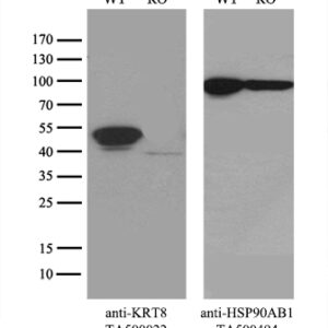
Imunógeno Fragmento de proteína recombinante humana correspondente aos aminoácidos 91-381 de CK8 humana (NP_002264) produzida em E. coli. Aplicações WB 1:500~1000, IHC 1:50, IF 1:50~100 Aplicações2 WB, IHC, IF Resumo A queratina 8 é um membro da família da queratina tipo II agrupada no braço longo do cromossomo 12. As queratinas tipo I e tipo II se heteropolimerizam para formar filamentos de tamanho intermediário no citoplasma das células epiteliais. O produto desse gene tipicamente dimeriza com queratina 18 para formar um filamento intermediário em células epiteliais simples de camada única. Esta proteína desempenha um papel na manutenção da integridade estrutural celular e também atua na transdução de sinal e na diferenciação celular. Mutações neste gene causam cirrose criptogênica. Formulação PBS (pH 7.3) contendo 1% BSA, 50% glicerol e 0.02% azida sódica. Purificação Purificado a partir de fluidos de ascite de camundongo por cromatografia de afinidade Isotipo IgG1 Reatividade Humano, Macaco Hospedeiro Camundongo Tamanho 53.5 kDa Tipo UltraMAB Concentração 0.20 mg/ml -
 R$12.423,26
Imunógeno Proteína recombinante humana de comprimento total de HMBS humano (NP_000181) produzida na célula HEK293T. Aplicações WB 1:2000, IHC 1:200, Aplicações2 WB, IHC Resumo Este…